3D embryo atlas reveals human development in unprecedented detail
Digital model will aid vital research, offering chance chance to explore intricate changes occurring in the first weeks of life
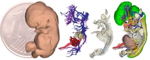
The beautiful and otherworldly development of the human embryo has been revealed in unprecedented detail in an interactive three-dimensional atlas.
The digital models, built by a team of scientists in the Netherlands, took around 45,000 hours to produce and offer researchers an unparalleled glimpse into the first eight weeks of human development.
“Everyone thinks we already know this, but I believe we know more about the moon than about our own development,” said Bernadette de Bakker, of the University of Amsterdam, who co-led the project with colleague Professor Antoon Moorman.
As de Bakker pointed out, many textbook depictions of embryo development are based on observations made many decades ago, and often contain details inferred from studies on mouse or chick embryos.
By contrast, the new resource will offer researchers the chance to explore the intricate changes occurring in the first weeks of life from a series of human specimens, aiding vital research. “It is important to understand normal human embryology to clarify how inborn defects and congenital malformations occur,” said de Bakker.
Published in the journal Science and available for all to view on a special website, the research involved the painstaking construction of 3D digital models of human embryos at various stages during the first two months of development.
Around 75 trained students were involved in analysing digital photographs of approximately 15,000 stained sections of tissues from the US-based Carnegie Collection of embryos – an array dating from around 60 to more than 100 years ago, collected by doctors during procedures such as hysterectomies.
For each section, organs and tissues had to be carefully labelled in order to build up the digital atlas. “If you look at one section, you have to draw a line around the liver, you have to draw a line around the kidneys, draw lines around the nerves, the blood vessels – and that is done manually by a digital pen and tablet,” said de Bakker, adding that up to 1,000 sections were manually analysed for each embryo, with as many as 150 organs and structures identified. In total, sections from 34 embryos were analysed, two for each of 17 different stages of development.
The resulting atlas is currently composed of interactive, 3D models of 14 of the embryos, each corresponding to a different stage within the first two months of development.
Using the tool, scientists can explore the development and changing position of different organs, as well as structures such as the skeleton or nervous system, rotate the embryo and select features of interest. Also available to view are the digital photographs of the sections used to produce the models, as well as detailed figures and tables of data containing unprecedented information on how each organ grows and changes over time.
The work has already yielded new insights. “We discovered that some organs in humans develop [far] earlier than they first arrive in chick or mouse embryos and some [develop far] later,” said de Bakker. That, she says, is important for areas such as developmental toxicology, where mouse or chick embryos are used as model organisms to explore the impact of various drugs or other chemicals.
SEE More here at the Guardian: https://www.theguardian.com/science/2016/nov/24/3d-embryo-atlas-reveals-human-development-in-unprecedented-detail


