Chromosomal Abnormalities
Remember that in meiosis, the genetic material separates into chromosomes. Chromosomal abnormalities are large-scale duplications or deletions of sections of chromosomes or entire chromosomes. The genetic material ends up “in the wrong place” influencing the development of the baby. Many times, the zygotes die at this stage and do not develop into embryos.
In babies carried to term, the most common chromosomal abnormalities are trisomy 21 (Down syndrome), trisomy 13 (Patau syndrome), and trisomy 18 (Edwards syndrome). Disabilities result directly from these chromosomal abnormalities. These are not considered environmental or multifactorial conditions.
Down Syndrome
How does Down Syndrome occur? What happens?
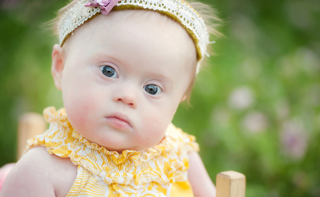
Down syndrome occurs when an individual has a full or partial extra copy of chromosome 21. This extra copy can originate either with the father (5% of cases) or the mother (95% of cases.) This additional genetic material alters the course of development and causes the characteristics associated with Down syndrome. A few of the common physical traits of Down syndrome are low muscle tone, small stature, an upward slant to the eyes, and a single deep crease across the center of the palm - although each person with Down syndrome is a unique individual and may possess these characteristics to different degrees, or not at all.

Down syndrome increases in frequency as a woman ages. However, due to higher birth rates in younger women, 80% of children with Down syndrome are born to women under 35 years of age.
There is no definitive scientific research that indicates that Down syndrome is caused by environmental factors or the parents' activities before or during pregnancy.
- One in every 550 babies in Ireland is born with Down syndrome, making Down syndrome the most common genetic condition.
- There are an estimated 5,000 Irish people with Down syndrome, and about 120 babies with Down syndrome are born in Ireland each year.
- In 1910, children with Down syndrome were expected to survive to age nine. With the discovery of antibiotics, the average survival age increased to 19 or 20.
- Now, with recent advancements in clinical treatment, most particularly corrective heart surgeries, as many as 80% of adults with Down syndrome reach age 60, and many live even longer.
- In 1866 English physician John Langdon Down, an English physician, published the first accurate picture of the person with Down syndrome. This earned Down the recognition as the “father” of the syndrome.
- In 1959, the French physician Jérôme Lejeune identified Down syndrome as a chromosomal condition. Instead of the usual 46 chromosomes present in each cell, Lejeune observed 47 in the cells of individuals with Down syndrome. It was later determined that an extra partial or whole copy of chromosome 21 results in the characteristics associated with Down syndrome.
- In the year 2000, an international team of scientists successfully identified and catalogued each of the approximately 329 genes on chromosome 21. This accomplishment opened the door to great advances in Down syndrome research.
Three types of Down syndrome
Types of Down syndrome are classified by how they originate. The most common type (95% of cases) is called Trisomy 21, which results in an embryo with three copies (trisomy) of chromosome 21 instead of the usual two. The pair of 21st chromosomes in either the sperm or the egg fails to separate. The extra chromosome is replicated in every cell of the body of the developing embryo. This brief video presents a simplified view:
In 1% of cases, some cells contain 46 chromosomes, and others 47, because only one of the initial cell divisions, rather than all of them, is involved. This type of Down syndrome is called mosaicism. Again, in the cells with 47 chromosomes, there will be an extra 21st chromosome present. Mosaicism may lead to fewer characteristics of Down syndrome.
In 4% of Down syndrome cases, part of chromosome 21 breaks off during cell division and attaches to chromosome 14. The baby has 46 chromosomes in the cell, but the presence of an extra part of chromosome 21 causes Down syndrome characteristics. This type of Down syndrome is known as translocation. If the father is the carrier, there is a 3% chance of another child being born with this syndrome; 10-15% if the mother is the carrier. And maternal age does not predict this form of Down syndrome. [1]
[1] National Down Syndrome Society (2012). ‘What is Down Syndrome?.’ Retrieved at http://www.ndss.org/Down-Syndrome/What-Is-Down-Syndrome/
Health Aspects of Down Syndrome
- The incidence of congenital heart disease in children with Down syndrome is between 40-60 percent. Some heart defects can be left alone with careful monitoring while others require surgery to correct the problem. The most common type is Atrioventricular Septal Defects (AVSDs).
- Some gastrointestinal problems occur more often in children with Down syndrome. They may be more common in children with Down syndrome but they are able to be treated or managed, thus giving the child the prospect of a healthier future.
- Children with Down’s syndrome all have some degree of learning disability, and at birth it is impossible to tell what degree that is.
- Many children with Down’s syndrome also have some degree of deafness. Deafness that is not diagnosed or managed can have a negative effect on a child’s social and language development, especially if a child has a learning disability. The PDF download file at the bottom of this page explains about different types and causes of deafness, how hearing is tested, what the results of a hearing test mean, and how deafness can be managed or treated.
Due to health problems and developmental disabilities, older children with Down syndrome in the past typically did not participate fully in school activities with other children. Things have changed… as we see this team assistant suit up and score a basket in an eighth grade basketball game!
What is Trisomy 18?
- Trisomy 18 occurs in about 1 out of every 2,500 births. Many Trisomy 18 babies are stillborn.
- Trisomy 18 is more life-threatening in the early months and years of life than Down syndrome.
- Most infants with Trisomy 18 will be admitted to the intensive care unit. Baby boys are more likely to die in the neonatal period. Some babies will be able to go home with their families with nursing support.
- 10% of Trisomy 18 babies survive to their first birthday. A small number of adults (usually girls) are surviving into their 20’s and 30’s with developmental delays that require assisted living.
- Medical problems that Trisomy 18 babies may have include:
- VSD (Ventricular Septal Defect): a hole between the lower chambers of the heart
- ASD (Atrial Septal Defect): a hole between the upper chambers of the heart
- Coarctation of the aorta: a narrowing of the exit vessel from the heart
- Kidney problems
- Gastrointestinal problems, such as failure of the eosophagus to connect to the stomach, and the presence of the intestinal tract outside the stomach, and hernias
- Clenched hands, rocker bottom feet, low set ears, strawberry shaped head
- Pocket of fluid on the brain (choroid plexus cysts)
- Delayed growth and severe developmental delays
- Small jaw and small head [2]
This story about Caitlin, who has survived to date through her teenage years and is shown as a 7-8 year old girl in the video, gives a snapshot of how Trisomy children can progress from infancy to childhood:
[2] Trisomy 18 Foundation (2013). ‘What is Trisomy 18?’ Retrieved at http://www.trisomy18.org/site/PageServer?pagename=whatisT18_whatis
What is Trisomy 13?
Trisomy 13 (Patau syndrome), where an extra 13th chromosome is present, occurs in approximately 1 in 5,000 births. 80% of infants with Trisomy 13 syndrome will have a full trisomy (affecting all cells) while the remainder will have the translocation or mosaicism variety. It is similar to Trisomy 18 in many ways.
Physical features are distinct, and help doctors make an initial diagnosis that can be confirmed by genetic tests. Typical features include small head size, small eyes or an absent eye, faulty development of the retina, or cleft lip and palate. Less serious issues, such as variation in ear shape, palm of the hand, extra fingers and toes, changes to the heel, also alert doctors to the presence of Trisomy 13. This is important, because doctors need to check right away for life-threatening problems.
This baby boy was expected to die soon after birth. His journey through the first year of life is traced here. At the time of this writing, he is now three years old.
80% of Trisomy 13 children have congenital heart defects. Like Trisomy 21 and 18 babies, they may have openings between the chambers in the upper or lower chambers of the heart. In addition, they may suffer from patent ductus arteriosis, where a special foetal channel fails to close, impairing blood flow, and dextocardia, where the heart is actually located on the right side of the body.
Infant mortality in Trisomy 13 babies isn’t from these congenital heart defects. Rather, lung problems are more dangerous. Trisomy 13 babies often suffer from apnea (where breathing stops) or problems with lung development. Aspirational pneumonia is also a common danger, because these babies are prone to eosophageal reflux and feeding issues, which can cause them to aspirate food into the lungs.
This powerful video, “Choosing Thomas: Inside a family’s decision to let their son live” documents the short time an American couple had with their son Thomas, born with Trisomy 13:
Trisomy children who survive infancy remain medically fragile. They experience developmental and motor challenges as they grow older. Older children can have visual problems due to retinal issues, small eyes or absent eyes. Hearing loss is common. Slow growth, high blood pressure, seizures and scoliosis (curvature of the spine) are common.
30% of Trisomy babies have kidney defects. 60% have a rare brain defect called holoprosencephaly, where the forebrain fails to divide. Developmental disabilities are therefore common.[3]
Natalia, shown in this video, is a Trisomy 13 survivor. Her story illustrates the potential and challenges faced by these fragile children:
[1] SOFT (Support Organization for Trisomy), (2013). ‘What is Trisomy 13?’ Retrieved at http://trisomy.org/trisomy-13-facts/


