Your beating heart
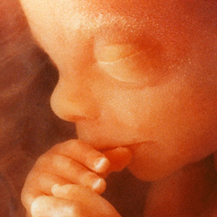
The most incredible organ in a young baby’s body is the heart. At the tender age of 21 days, the embryonic heart starts beating.
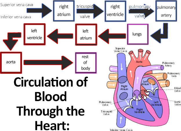
The circulatory process is a cycle involving the heart and the lungs. The heart exists to pump oxygenated blood through the body. We inhale oxygen through the lungs, and oxygenated blood travels to the heart through the pulmonary vein. The heart pumps this oxygen rich blood to the rest of the body through arteries and little capillaries. When the tissues have absorbed the nutrients from the blood, the capillaries and veins collect the depleted blood through the veins so it can return to the heart where it is re-oxygenated through the cardio-pulmonary cycle.
An unborn child cannot use her lungs to breathe in oxygen because there is no outside air inside the uterus.
The placenta functions as the 'lung'
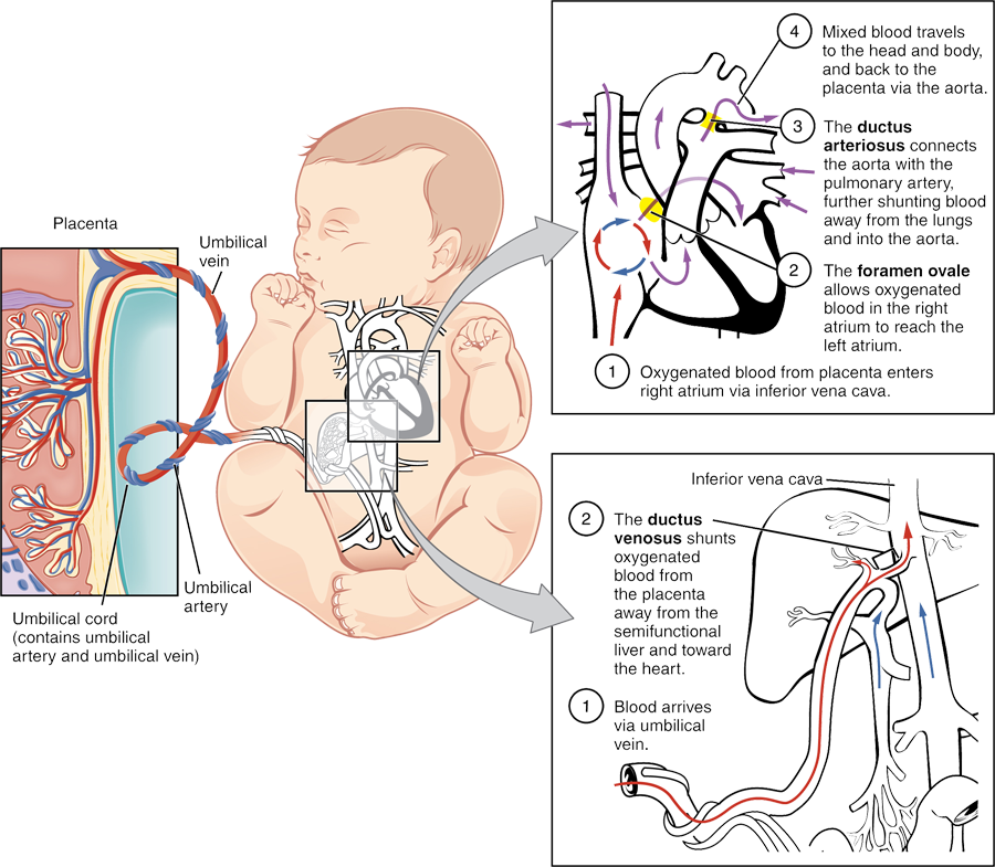
First, the placenta itself functions as the “lung” of the baby. Through the umbilical artery, blood comes in and then fills the placenta. Oxygen travels in (along with water, sugars, amino acids, and nutrients) and wastes leave through the placenta. But how does this 'lung' connect to the growing foetus?
The placenta is connected to its own “alveoli” (where oxygen and gas wastes are exchanged, as in the lungs.) This alveolus is called the chorionic villus, and it is connected to the umbilical vein, which carries the blood to the foetus.
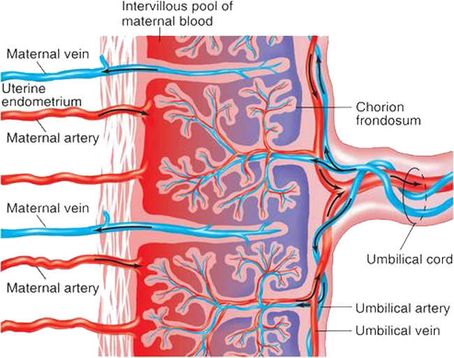
A portion of this blood enters the ductus venosus, which carries the blood to the inferior vena cava, which nourishes the lower part of the body.
The rest enters the liver, through the portal vein, and then the blood moves to the heart’s right atrium. The foetal heart has a hole between the upper atria called the foramen ovale. The blood passes from the right to left atrium without traveling down the right side of the heart, through the pulmonary system and back into the left side of the heart. This is because the placenta is the baby’s lung, and the blood is already oxygenated - the body is designed to use that lung and not the developing lungs inside the chest.
From there, most of the blood travels to the left ventricle of the heart, where it meets the aorta, which pumps the blood through the body. Some travels through the abdominal iliac arteries back to the placenta through the umbilical artery, and carries wastes out of the baby’s body.
But some blood does not enter the foramen ovale; it goes in the normal way through the right ventricle to the pulmonary artery. In an adult, the pulmonary artery would take the blood to the lungs, but there’s no need in the foetus. The foetal artery instead connects directly to the aorta, the main highway taking oxygenated blood to the body.
When you take your first breath...
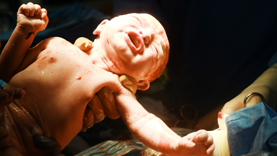
When the baby leaves the comfortable uterine “lung” and the umbilical cord is broken at birth, something incredible happens. Baby draws her first breath, and this inflow of air changes the pressure in the veins and arteries connecting heart and lungs, and in turn, changes the amount of pressure in the left atrium. There is then more pressure in the left atrium than in the right, and the little foramen ovale that connected the two chambers closes. The heart now has four distinct chambers, and blood flows from left atrium to left ventricle, through the pulmonary cycle, into the right ventricle and out through the right atrium. T
What about the ductus arterious, which kept blood away from the lungs? Well, when baby breathes, she takes in oxygen, and this in turn causes hormones called prostaglandins to decrease. The hormonal changes close the ductus arteriosus, and blood cannot escape past the pulmonary system anymore.
References and Sources
- Shier, D., et al. “The Cardiovascular System,” Hole’s Human Anatomy and Physiology, 13th edition. New York: McGraw-Hill, 2013. Pages 554-574.
- Course notes from Anatomy and Physiology II, Rosario Murdie, M.D. Ivy Tech State Community College North Central. 2013. Private collection accessed 30 August 2014.
- 1st Diagram - CNX.org - http://cnx.org/contents/29b65785-b2c5-4bec-9101-944f05f3c6af@3. Used under Creative Commons 4.0
- 2nd Diagram - Wikipedia - http://en.wikipedia.org/wiki/File:Circulation_of_Blood_Through_the_Heart.jpg Used under Creative Commons Licence. Date 3 February 2009
- 3rd Diagram - Human Physiology 2011 - http://humanphysiology2011.wikispaces.com/file/view/The_circulation_of_blood_within_the_placenta.jpg/222371454/493x410/The_circulation_of_blood_within_the_placenta.jpg


